Researchers Use Silkworm Silk to Model Muscle Tissue
By Anessa Pennington |

Researchers from the Biological Engineering and Biology Departments developed a new method of growing skeletal muscle cells: not on a plastic surface but on the three-dimensional silkworm silk fibers.
Researchers at Utah State University are using silkworm silk to grow skeletal muscle cells, improving on traditional methods of cell culture and hopefully leading to better treatments for muscle atrophy.
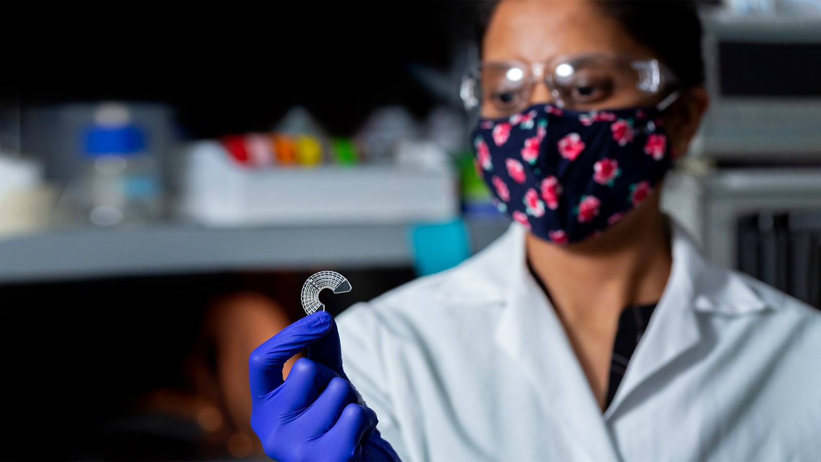
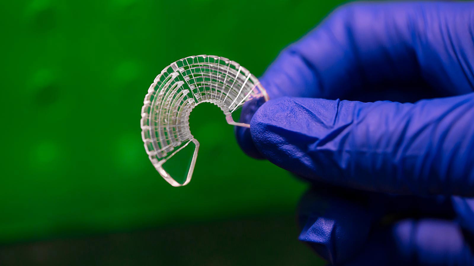
When scientists are trying to understand disease and test treatments, they generally grow model cells on a flat plastic surface (think petri dish). But growing cells on a two-dimensional surface has its limitations, primarily because muscle tissue is three-dimensional. Thus, USU researchers developed a three-dimensional cell culture surface by growing cells on silk fibers that are wrapped around an acrylic chassis. The team used both native and transgenic silkworm silk, the latter produced by silkworms modified with spider silk genes.
Native silkworm silks have been used previously as three-dimensional cell culture models, but this is the first time that transgenic silkworm silk has been used for skeletal muscle modeling. Elizabeth Vargis, Matthew Clegg and Jacob Barney of the Biological Engineering Department, and Justin Jones, Thomas Harris and Xiaoli Zhang of the Biology Department published their findings in ACS Biomaterials Science & Engineering.
Cells grown on silkworm silk proved to more closely mimic human skeletal muscle than those grown on the usual plastic surface. These cells showed increased mechanical flexibility and increased expression of genes required for muscle contraction. Silkworm silk also encouraged proper muscle fiber alignment, a necessary element for robust muscle modeling.
Skeletal muscle is responsible for moving the skeleton, stabilizing joints, and protecting internal organs. The deterioration of these muscles can happen for myriad reasons, and it can happen swiftly. For example, after only two weeks of immobilization, a person can lose almost a quarter of their quadricep muscle strength. Understanding how muscles can atrophy so quickly must begin at a cellular level, with cells grown to better represent reality.
“The overarching goal of my research is to build better in vitro models,” said Elizabeth Vargis, associate professor of biological engineering at USU. “Researchers grow cells on these 2D platforms, which aren’t super realistic, but give us a lot of information. Based on those results, they usually transition into an animal model, then they move onto clinical trials, where a vast majority of them fail. I’m trying to add to that first step by developing more realistic in vitro models of normal and diseased tissue.”

Silk fibers are wound around an acrylic chassis to produce a three-dimensional cell culture device. Skeletal muscle cells grown on silkworm silk proved to more closely mimic human skeletal muscle than those grown on the usual plastic surface. Photo: Matt Jensen.
WRITER
Anessa Pennington
Public Relations Specialist
College of Engineering
435-797-7512
anessa.pennington@usu.edu
CONTACT
Elizabeth Vargis
Biological Engineering
Associate Professor
435-797-0618
elizabeth.vargis@usu.edu
Justin Jones
Assistant Professor
Department of Biology
435-797-9292
justin.a.jones@usu.edu
TOPICS
Research 875stories Engineering 334stories Health 305stories Biology 162stories Biotechnology 25storiesComments and questions regarding this article may be directed to the contact person listed on this page.


